CALL OUR SALES TEAM FOR A QUOTE TODAY
Product information “Cubital Fossa”
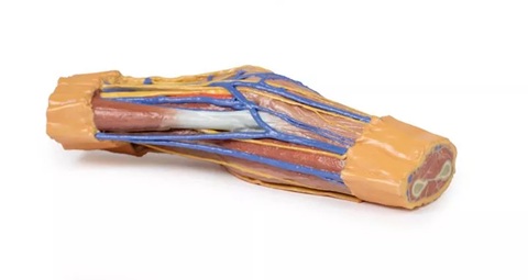
This 3D printed cubital fossa presents a superficial dissection of the right distal arm and proximal forearm.
The skin and superficial fascia have been removed anteriorly, medially, and laterally to reveal the superficial veins—basilic, cephalic, and median cubital—as well as the cutaneous nerves (medial, lateral, and posterior antebrachial).
Deep Fascia and Connective Tissue
The deep fascia underlying the superficial structures has largely been removed, but the antebrachial fascia is retained medially to demonstrate the merging of connective tissue fibers with the tendon of the biceps brachii through the bicipital aponeurosis. Medially, the ulnar artery is visible entering the cubital tunnel proximal to the medial epicondyle of the humerus. Anteriorly, the median nerve, brachial artery, and accompanying veins run parallel to the biceps brachii. On the lateral aspect, the cephalic vein rests on the brachioradialis muscle, while the posterior antebrachial cutaneous nerve lies on the common origin of the forearm extensors, just anterior to the exposed origin of the triceps brachii.
Cross-Sectional Anatomy
The proximal cross-section displays the anterior and posterior arm compartment muscles (biceps brachii, brachialis, triceps brachii), neurovascular bundles (median, ulnar, and radial nerves; brachial artery and veins), and superficial veins (basilic and cephalic) at the midshaft of the humerus.
The distal cross-section reveals the anterior and posterior forearm compartment muscles separated by the interosseous membrane, along with superficial and deep neurovascular bundles (radial artery, vein, and superficial branch of the radial nerve; ulnar artery, vein, and nerve; median nerve; anterior and posterior interosseous arteries, veins, and nerves) and the distal continuations of the superficial veins and cutaneous nerves.