25 Microscope Slides.
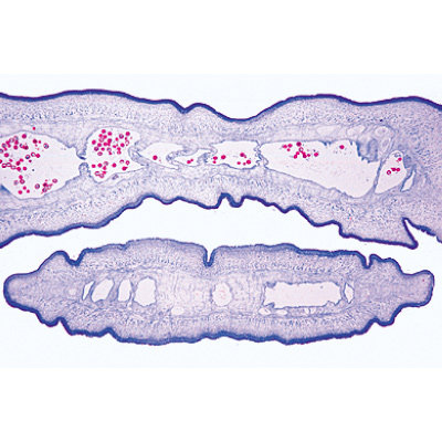
1(f) Trypanosoma gambiense, Central African sleeping disease, blood smear 2(f) Plasmodium berghei, malaria in rodents, blood smear with vegetative forms and schizogony stages 3(f) Sarcocystis sp., section of muscle showing the parasites in Miescher’s tubes 4(e) Nosema apis, honey bee dysentery, t.s. of diseased bee intestine 5(d) Eimeria stiedae, causes coocidiosis in rabbit liver, t.s. shows parasites in all stages 6(f) Fasciola hepatica, beef liver fluke, w.m. of adult flat mount and carefully stained 7(d) Fasciola hepatica, ova w.m. 8(t) Taenia or Moniezia, tapeworm, scolex w.m. 9(f) Taenia pisiformis, dog tapeworm, mature proglottids w.m. 10(d) Taenia saginata, tapeworm, proglottids in different stages t.s. 11(f) Hymenolepis nana, dwarf tapeworm, proglottids w.m. 12(f) Echinococcus granulosus, cyst wall and scolices sec. 13(d) Ascaris lumbricoides, roundworm of human, adult female t.s. in region of gonads 14(d) Ascaris lumbricoides, ova from faeces w.m. 15(f) Enterobius vermicularis (Oxyuris), pin worm, adult specimen w.m. 16(d) Trichinella spiralis, muscle with encysted larvae l.s. 17(g) Ixodes sp., tick, adult w.m. Carrier of relapsing fever and borreliosis 18(d) Dermanyssus gallinae, chicken mite w.m. 19(e) Acarapis woodi, varroa, parasitic mite of honey bee, w.m. 20(e) Sarcoptes scabiei (Acarus siro), section of diseased skin with parasites 21(f) Anopheles, malaria mosquito, head and mouth parts of female w.m. 22(e) Culex pipiens, common mosquito, head and mouth parts of female w.m. 23(f) Cimex lectularius, bed bug, w.m. 24(f) Pediculus humanus, human louse, w.m. 25(e) Ctenocephalus canis, dog flea, adult w.m.


