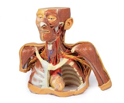
Head, Neck and Shoulder with angiosomes
CALL OUR SALES TEAM TODAY FOR YOUR REQUIREMENTS
Head and Neck:
The calotte has been removed ~2?cm above the orbits to expose the brain and endocranial cavity. A transverse cerebral section shows grey and white matter, lateral ventricles, and choroid plexus. The right side retains skin and fascia, false-coloured to highlight facial and neck angiosomes. The left side reveals facial expression and mastication muscles, and infratemporal structures including the lingual nerve and terminal branches of the external carotid artery. The carotid sheaths are opened bilaterally, exposing the common, internal, and external carotid arteries, and vagus nerves. The sternocleidomastoid and internal jugular veins are mostly removed. On the right, the great auricular and hypoglossal nerves are visible, along with the stylohyoid ligament and supra-/infrahyoid muscles. The thyroid gland is prominent, with preserved superior and inferior thyroid vessels.
Root of the Neck and Axilla:
On the left, partial clavicle removal reveals the first rib, anterior scalene, and brachial plexus roots (C5–T1) forming trunks between scalene muscles. The subclavian artery passes posterior to scalenus anterior, transitioning to the axillary artery, closely related to the brachial plexus cords.
The left axilla displays brachial plexus divisions and cords. The formation of the median nerve around the axillary artery is distinct. The ulnar, musculocutaneous, axillary, thoracodorsal, and long thoracic nerves are clearly identified with their courses and muscular targets.
On the right, the clavicle and subclavius muscle are intact, showing the cervico-axillary canal. Pectoralis major and minor have been reflected, exposing deeper structures.
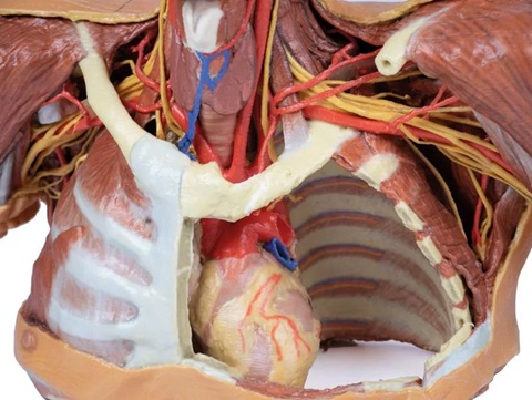
Thorax:
A window in the left thoracic wall reveals the mediastinum. The left lung has been removed. Intercostal spaces are visible beneath the parietal pleura; neurovascular bundles are identifiable posteriorly. The heart is exposed without pericardium, showing the left atrium and ventricle, pulmonary vessels, aorta, and both left vagus and recurrent laryngeal nerves. The right thoracic wall remains intact, displaying intercostal and upper limb muscles. From below, the right lung, pleural cavities, and diaphragmatic heart surface are visible. Posterior thoracic skin and fascia are intact, showing cutaneous nerve distribution.
Size: 50 x 20 x 41 cm