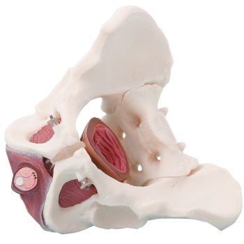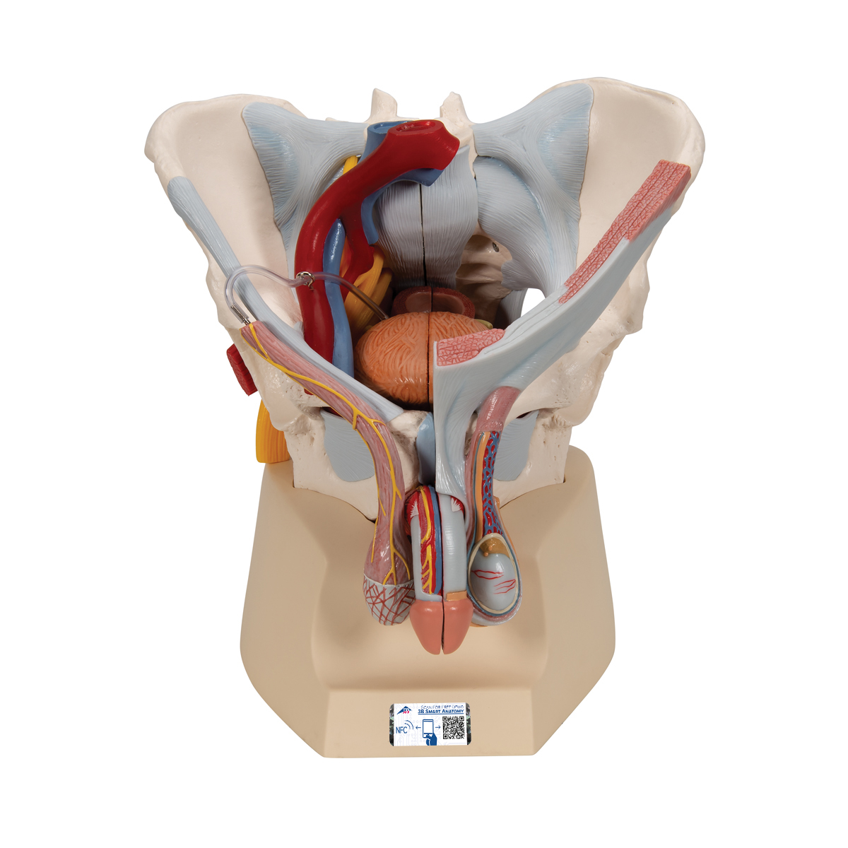
This 7 part model of the male pelvis shows in accurate detail how the bones, ligaments, vessels and nerves as well as the pelvic floor muscles and the external sex organs are connected to each other. It shows the whole pelvis, through which a median section has been placed. The right side of the external anal sphincter, the M. ischiocavernosus, the M. transversus perinei profundus and superficialis and the M. bulbospongiosus can be removed together.
The rectum, bladder, prostate and penis can also be removed, and split into two halves at median level – partially connected. The bone structures are connected with magnets and can therefore be easily taken apart. The skin and the fascia of the penis have been partly removed so that the vessels and nerves are visible. Part of the skin in the area of the scrotum and the spermatic cord has removed too. The testicles and epididymis are also visible. On the left, the spermatic cord has been opened up in layers, and on the right the M. cremaster and fascia spermatica interna have been exposed.
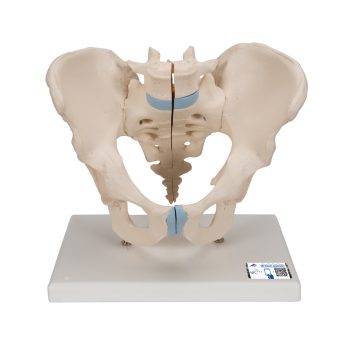
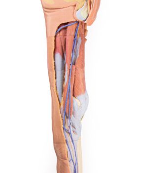
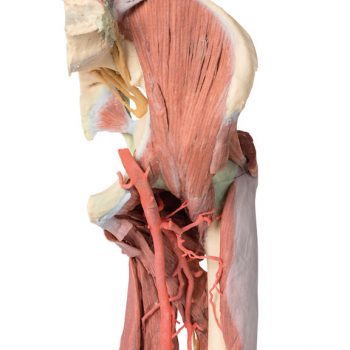
The right half of the pelvis shows sections of the common, external and internal iliac arteries and the common and external iliac veins, demonstrating their positions in relation to each other. The Plexus sacralis, the N. ischiadicus, the N. pudendus, the N. dorsalis penis, the Nn. scrotales anteriores, the Nn. perineales and the Nn. annales inferiores as well as the Ductus deferens are shown. The model shows the following bones and ligaments: both hip bones, pubic symphisis, sacrum and coccyx as well as the fifth lumbar vertebra with intervertebral disc. A median section has been placed through the fifth lumbar vertebra, the sacrum and the coccyx, so that the pelvis can be split into two halves. This means that part of the cauda equina is also visible in the vertebral canal. The left half of the fifth lumbar vertebra can be removed.
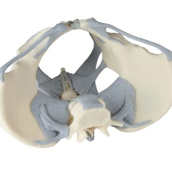
The following ligament structures of the pelvis are shown:
Lig. inguinale
Lig. sacrotuberale
Lig. sacrospinale
Lig. sacroiliaca anteriora
Lig. iliolumbale
Lig. longitudinale anterius
Lig. Supraspinale, Lig. sacroiliacum interosseum
Lig. sacroiliacum posterius
Lig. sacrococcygeum laterale
Lig. sacrococcygeum posterius superficiale et profundum
Membrana obturatoria
Lig. lacunare