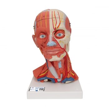
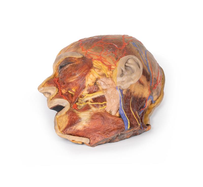
Product information “Superficial Facial nerves & Parotid Gland”
This 3D model presents the superficial anatomy of the face and head, and compliments the superficial facial anatomy of our HW 44 model with a more expanded dissection across the scalp and occipital regions.
The superficial neurovascular and muscular structures in the face largely mirror the structures described in reference to our HW 44 specimen (see description), although the terminal branches of the facial nerve (CNVII) can be largely followed across a longer course from the parotid gland and the platysma muscle has been retained superficial to the mandible and extends towards the neck.
In contrast to the HW 44 specimen, this model has a more expansive superficial dissection inferior to the external ear and across the posterior scalp and occipital region. This allows for an expanded appreciation of the neurovascular distribution of the supraorbital and supratrochlear nerves and arties with the superficial temporal artery. Inferior to the ear, the retromandibular vein has been exposed with the ascending fibres of the great auricular nerve on its superficial surface (and further branches of this nerve on the surface of the sternocleidomastoid muscle). At the posterior border of the sternocleidomastoid muscle the lesser occipital nerve is just preserved, near the exiting and ascension of the occipital artery and vein near the trapezius muscle towards the posterior scalp. Surrounding the external ear are fibres of the auricularis superior and posterior muscles. Near the margin of the dissection window posteriorly the deep fibres of the occiptalis muscle can be seen integrated into the epicranius (occipitofrontalis) muscle.
In Stock
Superficial Facial nerves & Parotid Gland”
€1,699.00